Cusabio Cell cycle Recombinants

What is the cell cycle?

The cell cycle, also called the cell division cycle, refers to the process from the completion of the division of a continuously dividing cell to the completion of the next division. In this biochemical process, a large amount of DNA in a cell’s chromosome is precisely duplicated, and then the copies are split exactly into two genetically identical daughter cells.

The function of the cell cycle

The cell cycle Recombinants is an important process by which a single-celled fertilized egg develops into a mature organism. And the cell cycle maintains and ensures the regeneration of hair, skin, blood cells and some internal organs.

The cell cycle process

In eukaryotic cells or cells with a nucleus, the phases of the cell cycle include two main stages: the interphase and the mitotic (M) phase.

1. Interphase:

Interphase is the duration between the end of a cell’s last division and the beginning of its next division. Interphase is an important period during mitosis. And it prepares for cell division and makes the next mitosis possible. Many events take place during interphases, such as DNA replication, the synthesis of related proteins, and the gradual disappearance of the nuclear membrane and nucleoli. And interphase time accounts for about 91 per cent of cell division. Interphase comprises Gap 1 (G1) phase, Synthesis (S) phase, Gap 2 (G2) phase.

Sometimes, some cells drop out of the cycle and stop dividing under adverse conditions, such as nutrient deprivation. These cells enter a resting phase, the G0 phase. Among these cells, some could re-enter the cycle when given the right conditions, but some cells that do not proliferate can no longer divide like neuron cells: they have reached their final stage of development. So the cells that reside in the G0 phase can be a temporary or permanent rest.

G1: Also called the growth phase. In the duration of the G1 phase, the organelles enlarge and mRNA and protein are synthesized in the cells. And the cells get bigger. All of these alterations are primed for DNA synthesis. The checkpoint (restriction point) in the G1 phase determines whether a cell continues to divide or exits the cycle (enters the G0 phase). If a cell successfully passes through the G1 checkpoint, it acquires the admission ticket to continue dividing. The checkpoint is regulated by cyclin G1/S, which promotes the transition of cells from the G1 to the S phase.

S: The S phase completes the synthesis of DNA and histones that are involved in DNA assembly and chromatin composition. During this period, the amount of DNA doubles and each chromosome is copied into two chromatids that are joined at the centromere.

G2: The G2 phase is the gap between the end of DNA replication and the beginning of mitosis. Cells synthesize certain proteins and RNA molecules to provide material to enter mitosis during the G2 phase. The G2 checkpoint, which is regulated primarily by the tumour protein p53, examines the cell for DNA damage within the chromosome before the cell enters the mitotic phase. Once it traces DNA damage, p53 can either repair DNA or trigger apoptosis. If p53 mutates or malfunctions, DNA-damaged cells can continue through the cell cycle, potentially leading to the development of cancer.

2. Mitotic Phase (M):

Mitosis occurs only in eukaryotes. Prokaryotes divide by binary division due to the absence of nuclei. The M phase is complex and highly regulated. The sequence of events is divided into prophase (including prophase and prometaphase), metaphase, anaphase, and telophase. Mitosis accounts for about 10 per cent of the cell cycle (it can last only an hour or two) and is much shorter than interphase. Mitotic errors can cause cell death by apoptosis or mutations that can induce cancer.

Prophase: During prophase, the nuclear envelope disintegrates and the nucleolus disappears. Chromatin condenses into coils to form chromosomes. And the centrosome emits star rays (in animal cells) or the cell poles send out spindle filaments (in plant cells) to form spindles. And then the spindle fibres attach to the centromere of the chromosome.

Metaphase: When the spindle fibres attach to the centromeres, they pull the chromosomes toward the centre of the cells, where all the chromosomes line up at the surface of the equator.

Anaphase: In this stage, the centromere splits and the two sister chromatids are attracted to two poles of the cells by the spindle fibre. As a result, the number of chromosomes doubles.

Telophase: the nuclear envelope reforms and the nucleolus appears. Also, the chromosomes gradually unzip to form chromatin, and the spindles gradually disappear. Subsequently, cytokinesis occurs, which divides the nucleus, cytoplasm, organelles, and cell membrane into two daughter cells that contain nearly equal parts of the parent cell.

Cytokinesis works differently in animal cells and plant cells. In animal cells, a protein near the equator forms the contraction ring to pinch the cell in half, creating shallow grooves on the surface. Due to cell walls, plant cells do not form shrinking rings but instead, build cell plates in the middle of the cells and then regenerate new cell walls in the two daughter cells.

Regulation of the Eukaryotic Cell Cycle

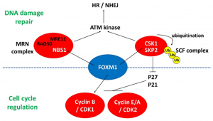
Regulation of the eukaryotic cell cycle, including detection and repair of DNA damage and prevention of uncontrolled cell division, is critical for cell survival. The molecular events that control the cell cycle are ordered and directed. The completion of the cell cycle process depends on the precise and strict regulation of the cell cycle by various regulatory factors. The core of these regulatory factors is:

Cyclin-dependent kinase (CDK): Cyclin-dependent kinase inhibitor (CKI) is a negative regulatory factor for CDK, while cyclin can upregulate CDK.

MPF (M phase promoting factor): It is a factor that can induce interphase cells to enter the division stage early in the M ​​phase cells of all eukaryotes. MPF can catalyze the CDK subunit so that it remains constant in amount. And it is regulated by Cyclin. MFP accumulates and is broken down at different phases of the cell cycle.

Cellular Checkpoints: Primarily detect whether early cell cycle events have been completed and cells are intact, and monitor DNA damage or delay responses during cell cycle progression. Here are some cell checkpoints listed below.

● The G1/S checkpoint is the rate-limiting step in the cell cycle. It is responsible for checking if the cells have enough material to fully replicate the DNA (nucleotide bases, DNA synthase, chromatin, etc.).

● The S checkpoint checks whether DNA replication is complete.

● The G 2 / M checkpoint is the checkpoint where the cell makes sure it has enough cytoplasm and phospholipids to divide the cell in two. Sometimes it checks if the replication time is correct.

● The metaphase-anaphase checkpoint is the test point of the spindle assembly. Failure during the attachment of centromeres to the spindle inhibits APC activity, resulting in cell cycle arrest.

Cell Cycle and Diseases

The cell cycle is associated with a variety of human diseases, especially cancer. Uncontrolled cell proliferation caused by cell cycle disorders is the main cause of cancer. At the molecular level, it is the result of genetic mutations that cause inappropriate activation of cell cycle promoters and/or deactivation of inhibitors, resulting in uncontrolled cell cycle regulation.

Related Cell Cycle Applications

Recently, some experts have designed some drugs that aim to stop cell spindle formation and further inhibit cell mitosis and maintain cell division in the G0 phase. The drugs effectively slow down malignant proliferation and the spread of cancer cells.

For example, ordinary watermelons are diploid and produce normal seeds. Ordinary watermelons treated with colchicine can make tetraploid watermelons and produce normal seeds. Due to colchicine’s inhibition of spindle formation, mitosis is repressed and chromosomes are arrested at the metaphase of division. In such mitosis, the chromosomes divide longitudinally, but the cells do not divide and cannot form two daughter cells, so the chromosomes are duplicated. When a diploid watermelon is crossed with a tetraploid watermelon, the triploid watermelon is produced. Since triploid watermelon cannot distribute chromosomes equally among gametes, normal seeds cannot be obtained. This is a seedless watermelon

Cusabio C-terminal GST-tagged Recombinant

Abstract

C-terminal Glutathione S-transferase (GST) tagged Recombinant from Schistosoma japonicum, which is widely used for the production of fusion proteins in the cytoplasm of Escherichia coli, was employed as a functional fusion cassette that affects dimer formation of a recombinant protein and confers indicator enzyme activity at the same time. For this purpose, GST was linked via a flexible spacer to the C-terminus of the thiol-protease inhibitor, cystatin, whose papain-binding properties were to be studied.

The fusion protein was secreted into the bacterial periplasm via the OmpA signal peptide to ensure the formation of the two disulfide bonds in cystatin. The formation of erroneous crosslinks in the oxidizing medium was prevented by replacing three of the four exposed cysteine ​​residues in GST. Using the tetracycline promoter for tightly controlled gene expression, the soluble fusion protein could be isolated from the periplasmic protein fraction. Purification to homogeneity was achieved in a single step by means of a glutathione agarose affinity column.

Alternatively, the protein was isolated by streptavidin affinity chromatography after the Strep-tag had been added to its C-terminus. The GST moiety of the fusion protein was enzymatically active and kinetic parameters were determined using glutathione and 1-chloro- 2,4-dinitrobenzene as substrates. In addition, strong papain binding activity was detected in an ELISA. The signal with the cystatin-GST fusion protein was much higher than with cystatin itself, demonstrating an avidity effect due to the formation of GST dimers.

The quaternary structure was further confirmed by chemical crosslinking, which resulted in a specific reaction product with twice the molecular size. Therefore, the engineered GST is suitable as a secretion-competent fusion partner of moderate size that can confer bivalence to a protein of interest and promote detection of binding interactions even in low-affinity cases.

The GST label

Protein purification with affinity tags, such as glutathione S-transferase (GST), histidine (HIS) and other affinity tags, allows the purification of proteins with both known and unknown biochemical properties. Therefore, this methodology has become a widely used research tool to determine the biological function of uncharacterized proteins. GST is a 211 amino acid (26 kDa) protein whose DNA sequence is frequently integrated into expression vectors for recombinant protein production.

The result of expression from this vector is a GST-tagged fusion protein in which the functional GST protein (26 kDa) is fused to the N-terminus of the recombinant protein. Because GST folds rapidly into a stable and highly soluble protein upon translation, the inclusion of the GST tag often promotes greater expression and solubility of recombinant proteins than expression without the tag. Additionally, GST-tagged fusion proteins can be purified or detected based on the ability of GST (an enzyme) to bind to its substrate, glutathione (GSH).

GST fusion protein purification

Glutathione is a tripeptide (Glu-Cys-Gly) that is the specific substrate for glutathione S-transferase (GST). When reduced glutathione is immobilized via its sulfhydryl group on a solid support, such as cross-linked bead agarose, it can be used to capture pure GST or GST-tagged proteins through enzyme-substrate binding reaction.

Immobilized Glutathione

Binding is most efficient in near-neutral buffers (physiological conditions) such as Tris-buffered saline (TBS) pH 7.5. Because binding depends on the preservation of the essential structure and enzymatic function of GST, protein denaturants are not compatible.

After washing an affinity column to remove unbound sample components, the purified GST fusion protein can be dissociated and recovered (eluted) from a glutathione column by the addition of excess reduced glutathione. Free glutathione competitively displaces the binding interaction of immobilized glutathione with GST, allowing the fusion protein to emerge from the affinity column.

This affinity system typically produces greater than 90% pure GST-tagged recombinant protein from crude bacterial or mammalian cell lysate samples. Glutathione-based affinity purification of GST-tagged fusion proteins is easily performed on a small, medium, or large scale to produce microgram, milligram, or gram quantities.

At 26 kDa, GST is considerably larger than many other fusion protein affinity tags. For reasons that have not been fully characterized in the literature, the GST fusion tag structure is often degraded upon denaturation and reduction for protein gel electrophoresis (eg, SDS-PAGE). As a result, electrophoresed samples of GST fusion proteins often appear as a ladder of lower MW bands below the full-size fusion protein.

Cusabio Macaca mulatta Recombinant

Bottom

Scrub typhus is a major endemic disease in tropical Asia caused by Orientia tsutsugamushi for which an effective, broadly protective vaccine is not available. Successful evaluation of candidate vaccines requires well-characterized animal models and a better understanding of the immune response against O. tsutsugamushi. While many animal species have been used to study host immunity and vaccine responses in scrub typhus, only limited data exist in non-human primate (NHP) models.

Findings of methodology/principles

In this study, we evaluated an NHP scrub typhus disease model based on intradermal inoculation of the Karp strain of O. tsutsugamushi in Macaca mulatta Recombinant (n = 7). After intradermal inoculation with 106 murine LD50 of O. tsutsugamushi in the anterior thigh (n = 4) or mock inoculum (n = 3), a series of chronological investigations including haematological, biochemical, molecular and molecular assays were performed. immunological, until day 28, when tissues were collected for pathology and immunohistochemistry.

In all NHP inoculated with O. tsutsugamushi, but not sham inoculated, the development of a classic eschar with central necrosis, regional lymphadenopathy, and elevated body temperature was observed on days 7 to 21 post-inoculation (pi). ; bacteremia was detected by qPCR on days 6-18 pi; and impaired liver enzyme function and increased white blood cells on day 14 pi. Immunological assays demonstrated elevated serum levels of soluble cell adhesion molecules, anti-O. Tsutsugamushi-specific antibody responses (IgM and IgG) and pathogen-specific cell-mediated immune responses in inoculated macaques. qPCR assays detected O. tsutsugamushi in eschar, spleen, draining and non-draining lymph nodes, and double immunostaining demonstrated intracellular O. tsutsugamushi in eschar and lymph node antigen-presenting cells.

Product name

(Macaca Rhesus) Odontogenic Ameloblast Associated Protein (ODAM), Recombinant Protein

Full product name

Recombinant Macaca mulatta (Rhesus Macaque) Odontogenic Ameloblast Associated Protein (ODAM)

Product Synonym Names

Recombinant odontogenic ameloblast associated protein (ODAM) (Rhesus macaque); Odontogenic ameloblast associated protein; apin

Product gene name

ODAM Recombinant Protein

Product Synonym Gene Name

ODAM; APIN

For research use only

For research use only. It should not be used in diagnostic procedures.

Chromosome location

chromosome: 5; NC_007862.1 (59476282..59484317, plugin). Location: Chromosome: 5

3D structure: ModBase 3D structure for A1YQ92

Host: E Coli or Yeast or Baculovirus or Mammalian Cell

Purity/Purification

Greater than or equal to 85% purity as determined by SDS-PAGE. (lot specific)

Form/Format

Lyophilized or liquid (Format to be determined during the manufacturing process)

Label information

This protein contains an N-terminal tag and may also contain a C-terminal tag. The types of tags are determined by several factors, including the stability of the tagged protein; ask for information about the labels.

Sterility: Sterile filter available on request.

Endotoxin: Low endotoxin available on request.

Species: Mulatto macaque (Rhesus macaque)

Preparation and Storage

Store at -20 degrees C. For long-term storage, store at -20 or -80 degrees C.

ISO certification

Manufactured in an ISO 9001:2015 certified laboratory.

Other notes

On occasion, small volumes of ODAM recombinant protein vials may become trapped in the product vial seal during shipping and storage. If necessary, briefly spin the vial in a tabletop centrifuge to dislodge any liquid in the vial cap. Certain products may require dry ice shipping and an additional dry ice fee may apply.

Precautions

All MyBioSource products are for scientific laboratory research purposes and not for diagnostic, therapeutic, prophylactic, or in vivo use. Through your purchase, you expressly represent and warrant to MyBioSource that you will properly test and use any Product purchased from MyBioSource in accordance with industry standards. MyBioSource and its authorized distributors reserve the right to refuse to process any order where we reasonably believe the intended use will not meet our acceptable guidelines.

Disclaimer

While every effort has been made to ensure the accuracy of the information provided in this datasheet, MyBioSource shall not be responsible for any omissions or errors contained in this document. MyBioSource reserves the right to make changes to this datasheet at any time without notice.

Conclusions/Importance

These data show the potential of using rhesus macaques as a model of scrub typhus, for the evaluation of correlates of protection in both natural and vaccine-induced immunity and support the evaluation of future candidate bush typhus vaccines.