
Real-time PCR technology is becoming more and more popular in different industries. This technology is based on the detection and quantification of a fluorescent reporter whose emission is directly proportional to the amount of amplicons generated during the PCR reaction.
Since it generally uses closed tube systems and quantification requires no manipulation. Ostamplification, post-PCR contamination problems with amplicons are significantly reduced.
The entire process is automated from start to finish, making this technology very efficient for large-scale analysis applications. This article presents a description of the principles behind real-time PCR, the different amplicon detection technologies and examples of common applications.

Real time PCR, since when?
Russell Higuchi was one of the first to analyze kinetics of PCR (polymerase Chain Reaction) in developing a system that detected the PCR product at as it accumulated.
This system in “time real “was using ethidium bromide as an agent intercalating in each of the amplification reactions and a thermocycler modified to stimulate the emission of UV radiation samples. The broadcast of the fluorescence was detected using a CCD camera (charge-coupled device).
An increase in emission fluorescence was observed when bromide
of ethidium attached to the double stranded DNA produced during amplification. By tracing the increase in emission of fluorescence as a function of the number of cycles, the system produces amplification curves exhibiting a more complete diagram of the PCR process than the simple determination of amplicons (amplification products) accumulated at the end of amplification.
How does it work?
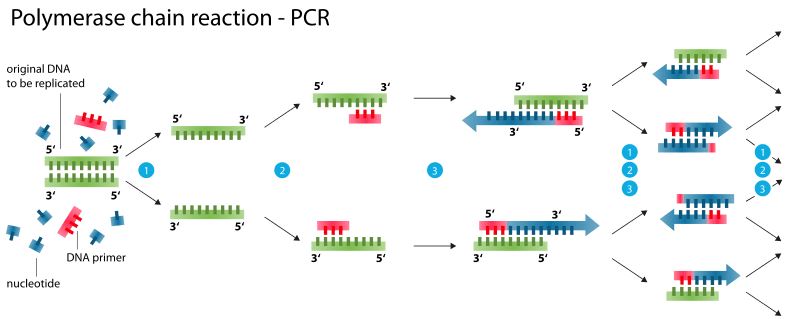
Since its invention, PCR has become the most no longer used for the detection of DNA and RNA from a simple copy of a particular sequence nucleic acids, this sequence can be specifically amplified and detected. His nature exponential makes this technique attractive for quantitative analyzes. Theoretically, there is a quantitative relationship between the quantity of the target sequence starting point and the quantity of the amplified product at any cycle. In practice, it is not uncommon for PCR reactions in replica give different levels of amplicons.
The development of real-time quantitative PCR a eliminated traditional variability associated with PCR quantitative and allows the quantification of the PCR product reliably and routinely. At the start of the PCR reaction, the reagents are in excess but in fairly low concentration to prevent the
renaturation of amplicons does not compete with primers hybridization. The amplification is then performed steadily at an exponential rate at
using thermostable DNA polymerase. After the phase exponential, the amplification reaction enters a linear phase where the amplification rate becomes extremely variable, even at the replica level of a same sample, due to competition between renaturation of amplicons and hybridization of primers. Then follows a plateau phase where the rate of amplification
decreases to almost zero generating very few amplicons.
In order to collect quantitative data accurately, each of the samples must be analyzed in its phase exponential amplification which is the most reproducible from the PCR reaction. Real-time PCR therefore monitors the fluorescence emitted during the reaction with an indicator of amplicon production during each cycle, as opposed to quantitative PCR conventional where amplicons are only detected at the end of the process.
Real-time PCR technology is based on the detection and quantification of a fluorescent “reporter”. The increase in fluorescent signal is directly proportional to the amount of amplicons generated during the PCR reaction. By observing the amount of fluorescence emitted in each cycle, it becomes possible to follow the PCR reaction during its exponential phase where the first significant increase in the amount of amplicons is in direct correlation with the initial amount of the original target matrix (template). Several real-time PCR instruments are currently on the market.
These devices generally use a closed tube system and the quantification does not require any post-amplification manipulation, which minimizes or liminates the problems of contamination by amplicons following the PCR reaction and reduces the analysis time. The entire process is therefore automated from start to finish, making this technology attractive for high-throughput analysis applications.
Detection technologies
All real-time PCR systems are therefore based on detection and quantification of a fluorescent emitter during the amplification process and the increase in fluorescent emission signal is directly proportional to the amount of amplicons produced during the reaction. There are two general principles for detection quantitative amplicons: agents binding to DNA double strand (eg SYBR Green I) and fluorescent probes.
For the latter category, there are currently four main technologies: hydrolysis of probes (Taqman assay), hybridization of 2 probes (HybProbes), tags Molecular Beacons and scorpion primers (Scorpion primers). According to Wittwer et al. (1997), these different detection technologies would have a equivalent sensitivity. However, these technologies have differences in specificity.
1) Agents binding to double stranded DNA
Molecules that bind to double-stranded DNA can be divided into two classes: intercalating agents like ethidium bromide (Higuchi et al, 1992), YO-PRO-1 , SYBR Green I and agents attaching to the furrow minor groove binders like Hoeschst 33258 . Their show fluorescent increases when linked to DNA double strand. To be used in a PCR reaction in In real time, these agents must meet two requirements: increase in fluorescence when linked to double stranded DNA and do not inhibit the PCR reaction.
The SYBR Green I, whose link mechanism is not well defined, is the most commonly used agent. Its advantages are that it is economical, easy to use and has more sensitivity than ethidium bromide without inhibiting the
amplification reaction.
During the PCR amplification reaction, the dye free in solution exhibits little fluorescence. During the stage elongation, an increase in fluorescence is associated with the amount of dye binding to double DNA budding strand. When monitored in real time, the increase of the fluorescence signal is observed during the polymerization and the fluorescent emission decreases completely when DNA is denatured in step next. Consequently, the fluorescence emission is measured at the end of each elongation step for each cycles by a reading system integrated into the Real-time PCR that tracks the increase in the amount of DNA amplified during the reaction.
SYBR Green I technology does not require no fluorescent probe but its specificity rests entirely on its primers. It does not require therefore no particular expertise for the design of fluorescent probes and is unaffected by mutations in target DNA that influence hybridization of probes specific. Since the SYBR Green I attaches to any double DNA molecule bit, this technology has a certain versatility since the same agent can be used to detect more of an amplification product in the same sequence reaction.
This technology also has certain disadvantages:
1) since full double stranded DNA emits signals, it becomes impossible during the reaction to ensure the specificity of the amplicons or to discriminate between the different amplicons in the case of multiplexing;
2) the bad mis-primering, often generating bands superfluous DNA observable on electrophoresis gel, may lead to false positives or an overestimation of the quantification;
3) the fluorescence emission can be biased by the molecular weight of DNA amplified by a longer amplicon which will fix more molecules fluorescent compared to a shorter amplicon in the same reaction .
2) Hydrolysis of probes
Taqman technology is activity-based 5′-exonuclease of Taq polymerase to hydrolyze a probe hybridized to its target sequence on the amplicon during the PCR hybridization / extension step. A emitting fluorochrome (reporter) (ex. FAM: 6-carboxyfluorocein) is attached to the 5 ’end of the probe
hybridization and its emission is inhibited by a second fluorochrome suppressor (quencher) present at the end 3 ’(eg TAMRA: 6-carboxy-tetramethyl-rhodamine).
When stimulated, the emitting fluorochrome transfers its energy to the neighboring suppressor fluorochrome by the FRET principle (fluorescence resonance energy transfer) which dissipates this energy in the form of heat rather than emitting fluorescence. Given that the 5′-exonuclease activity of Taq polymerase is specific to double stranded DNA, free probes in solution remain intact and no fluorescence is issued. During the hybridization step, the probe and the primers attach to their complementary sequences
respectively.
In the next step, Taq polymerase begins elongation of the new DNA strand from the primer until it meets the probe in its path hybridized that it displaces and hydrolyzes with its activity 5′-exonuclease. The eporter is then released from the suppressor environment allowing emission
fluorescence which increases with each cycle proportional to the hydrolysis rate of the probe. As Taq polymerase will hydrolyze the probe only when this is hybridized to its complementary sequence, the temperature conditions of the polymerization step must be adjusted to allow the probe to remain hybridized during this stage. The majority of probes have a dissociation temperature (Tm) around 70ºC or 5-10oC higher than the primers.
Therefore, the Taqman technology uses a combined step
hybridization and polymerization at 60-62ºC ensuring the hybridization and the stability of the probe during the extension. This also allows maximum 5-exonuclease activity of Taq polymerase but, the effectiveness of the activity of polymerization of the enzyme will be slightly reduced to this suboptimal temperature. For long amplicons, a longer hybridization / polymerization stage or an increase in the concentration of Mn2 + or Mg2 +
may be necessary to stabilize hybridization of the probe to its target sequence. Principles to be respected in the design of Taqman probes are also applicable to other linear probes and include as general rules:
1) a length of 20-40 nucleotides,
2) a G-C content varying from 40- 60%,
3) no repeat pattern,
4) no sequence allowing hybridization or overlap
with the primers,
5) an A, a C or a T at the end 5 ’ because a G suppresses the fluorescence of the transmitter even after cleavage and,
6) a higher Tm of 5 to 10 oC as the primers to ensure that they will hybridize before the primers and they will remain hybridized during the combined ybridization and polymerization .
Fluorescent probes have the advantage of compared to agents binding to DNA increased specificity and better multiplexing capacity. The specificity of hybridization between the fluorescent probe and its sequence of target DNA significantly reduces the emission of nonspecific fluorescence due to bad pairings or primer-dimers. Multiplex reactions can be developed by using separate emitting fluorochromes linked to different probes in the same PCR reaction.Taqman technology is however less efficient and less flexible than other real-time technologies for detection of specific mutations.
3) Hybridization of 2 probes
This technology is also known as HybProbes and is based on the use of two probes linear complementary to a target sequence for maximize signal specificity . A first probe, blocked at its 3 ’end in order to prevent its extension during the elongation stage, carries in 3 ’a donor fluorochrome (FITC) which produces green fluorescent light when excited by a light source. Its emission spectrum is more wider than that of the acceptor fluorochrome (Red 640 or Red 705) attached to the 5 ’end of a second probe. In solution, the two probes are free and separate.
Being given that energy transfer by the FRET principle depends on the distance between the two fluorochromes, it then only fluorescence background noise results green emitted by the donor fluorochrome. During the stage of hybridization, the two probes attach to their sequences respective targets located within 10 nucleotides in a spin-to-tail arrangement. The proximity of the two fluorochromes allows the transfer energy of green fluorescein by the FRET principle to the red acceptor fluorochrome and causes its emission fluorescent.
During the polymerization step, both probes independently return to solution which suppresses the emission of red fluorescence).
The increase in red fluorescence is proportional the amount of DNA synthesized during the PCR reaction and begins to decrease when the amount of amplicons products becomes significant enough to cause
an amplified DNA competition with hybridization simultaneous of the 2 probes on the target DNA. This technology therefore has great specificity and also allows great flexibility in the design of probes. In addition, as the probes are not hydrolyzed, they are reused at each cycle.
In addition to the fact that the target sequence must be located towards the 3 ’end of the amplicon and the Tm of the two probes should be similar, the general principles in the design of the Taqman probes are applicable for this technology.
4) Molecular beacons
A molecular tag is a DNA hybridization probe in the shape of a hairpin. The portion of the probe that compose the loop is complementary to the target sequence DNA. The trunk of the molecular beacon is formed by two arms with complementary sequences. A fluorescent transmitter (FAM, TAMRA,
TET, ROX) is attached to the end of one of the arms and a suppressor (quencher) (4- (4’-dimethylamino-phenylazo) – benzene: DABCYL) is attached to the end of the other arm.
The suppressor is a non-fluorescent chromophore which dissipates the energy of the emitting fluorochrome as heat by the FRET principle. Free probes adopt in solution a hairpin structure and the trunk holds the
arms together for effective suppression of the broadcast fluorescence. During the hybridization stage, when the probe encounters a sequence which is complementary to it, it adopts a transient conformation which forces the trunk to separate thus freeing the 2 arms. The probe then hybridizes preferentially to its target complementary sequence on the matrix. In this conformation, the fluorochrome transmitter is away from its restaurant suppressor as well the fluorescence emission that can be detected while the other molecular tags remain in structure hairpin (closed position). If the sequence of target DNA does not perfectly match(mismatch) to the sequence of the molecular tag no hybridization and, therefore, no fluorescent emission occurs.
This is mainly due to the thermodynamic properties of the structure of the molecular beacon which especially promote the formation of the hairpin except in the event of perfect hybridization of the sequences. The specificity of this technology is such
that it can detect sequence differences at nucleotide close, which can sometimes be difficult to achieve with linear probe technologies. It therefore constitutes a efficient technology for detection and screening at
large scale of SNPs (single nucleotide polymorphisms). The probes are designed to remain intact during the amplification reaction and have to re-hybridize to the target at each cycle to measure the constituent signal
thus another advantage compared to Taqman probes hydrolyzed at each cycle.
The most significant disadvantage associated with the use of molecular beacons is the design of hybridization probes. An optimal design of the trunk of molecular beacons is crucial. With an inadequate design, the trunk could adopt a different conformation which would distance the
fluorochrome emitting from the immediate environment of the suppressor thus resulting in a population of wrong probes suppressed and significant background noise. In return, if the hybridization forces of the trunk (dependent on the length and composition of sequences arms) are too important, they can interfere with tag hybridization molecular to its target complementary sequence and fluorescence emission. A denaturing profile
precise thermal of each of the molecular beacons must be established to determine the characteristics of dissociation (melting) of the arms.
Several variants of this technology such as Sunrise primers (now under the trade name Amplifluor TM hairpin primers), cyclicons and scorpion rimers have been proposed for detection specific nucleic acids in solutions
homogeneous.
5) Scorpion primers
The scorpion primers represent a variant of the molecular beacon technology. Fluorochromes and the probe (molecular beacon part) are integrated into the same irreversible amplicon during PCR amplification. The design of a scorpion primer / probe is practically similar to that of molecular beacons. Adding a molecule of hexethylene glycol (HEG), too
called blocker (stop), is necessary to prevent DNA polymerase replication of the molecular tag during the PCR reaction. The HEG is therefore located after the fluorochrome suppressor and is followed by a primer region.
The emitting fluorochrome carried in 5 ’from the beacon region molecular can be FAM or ROX and the suppressor is normally methyl red. The priming region of the scorpion therefore makes it possible to integrate the molecular beacon into the new amplicon during the PCR reaction. The loop hairpin is drawn in order to allow hybridization of the probe to its complementary sequence target located on the amplicon.
This hybridization forces the hairpin to change conformation thus allowing the emission of fluorescence. Thelwell et al report that technology primer / scorpion probe is generally more effective than Taqman technologies and molecular beacons, particularly in a PCR program with very short cycles.
Threshold cycle
The concept of the “threshold cycle” is the basis of a precise and reproducible quantification for techniques fluorescent in PCR. The fluorescence values are recorded during each cycle and represent the
amount of amplicons produced at a specific point in the reaction . The more templates there are amplify at the start of the PCR reaction, the lower the number of cycles required to reach a point where the signal fluorescence emission will be statistically and significantly higher than background noise .
This point is defined as the threshold cycle (Ct) and will always appear during the phase exponential amplification. Therefore, the
quantification is not affected by the exhaustion of one of the reagents like during the plateau phase which explains why the real time system is so reproducible. The Ct value can be translated into a quantitative result in
comparing it with the Ct values generated with known quantification matrices .
Applications
Real-time PCR, because of its ability to produce quick, specific and quantitative results, find more in addition to applications in different areas. Although this list is not exhaustive, here are the examples most common applications. Real-time PCR now allows you to perform finer studies of gene expression from tissues or cell lines as for quantitative analysis of the expression of different genes in studies of modulation of the cell cycle with tissues exposed to non-genotoxic carcinogens (cie TNO BIBRA), promoter analyzes in cell lines with the chloramphenicol acetyl transferase reporter gene , studies of modifications of gene expression in experiments drugs / responses, the study of mRNAs variants resulting from alternative splicing
(Vandenbroucke et al, 2001), etc. It can also allow to standardize the uantity of starting DNA and to evaluate the amount of mRNA at the end of the reaction in studies of in vitro transcription.
Real-time PCR is also a powerful tool for mutation analyzes such as SNPs and studies of large-scale genotyping as for the gene for estrogen receptor .
More and more tests using tag technology molecules are designed for detection and rapid quantification of viral pathogens, bacterial and parasitic. Vet et al (1999) have already proposed a multiplex system which allows the detection and simultaneous quantification of HIV-1, HIV-2 retroviruses, human T-cell lymphotrophic viruses type I and type II with a detection threshold of 10 genome copies and Poddar (1999) for the detection of adenovirus. Tests detection have also been developed for Salmonella with sensitivity of 2 CFU per real-time PCR reaction and 1 CFU / ml after 6 h enrichment for Escherichia coli O157: H7 from raw milk and apple juice samples . In parasites, a real-time PCR test has was developed for Toxoplasma gondii . Several laboratories are currently working on the
point many other diagnostic tests for different pathogens.
Traditionally, analyzing the number of copies of a plasmid to assess the genetic stability of collections of cells was done by Southern blot
(Southern blot). Real-time PCR now allows to perform these analyzes much faster and more precise. It can also be used in the evaluation the amount of contaminating chromosomal DNA or residual bacterial or mammalian in the production of recombinant proteins.
The analysis of the bio-distribution of a vector is an element important in evaluating the effectiveness of genetical therapy. Real-time PCR simplifies studies to assess the presence / absence of DNA or mRNA target in different tissues and helps determine the equilibrium conditions of the expressed mRNAs (steady-state
expressed mRNA).
Conclusion
According to the literature, the technique most frequently used so far is probe technology hydrolysis (Taqman assay) although its popularity is
mainly due to its commercial maturity. This technology has proven to be more effective than agents interleaving at the level of specificity and multiplexing. The development of detection technologies based on
molecular beacons brings even more specificity among other things for the detection of SNPs and genotyping analyzes compared to technologies
based on linear probes. Real-time PCR turns out very interesting for large analysis applications scale since the entire process is automated from start to finish.
Like this technique typically includes closed tube systems and
quantification requires no post-amplification manipulation, post-PCR contamination problems by the amplicons are greatly reduced.
Gene expression, cell response to different drugs (drug / response), detection of mutations, genotyping, detection and quantification
pathogens, DNA and RNA quantification, expression and distribution trials for therapy gene, quantifying the number of copies in a cell collection and ultimately DNA evaluation residual are, currently, some examples of increasingly widespread applications of PCR in time real. Real-time PCR is therefore a powerful tool and recent developments in different technologies detection suggest more applications innovative each other.